Medical education & research · United Kingdom
Turn medical imaging into physical anatomy.
Patient-specific 3D anatomical models created from medical imaging for education, teaching and research.
From image segmentation to high-resolution resin printing, the process is led by clinicians with expertise in medical imaging, machine learning and 3D model production.
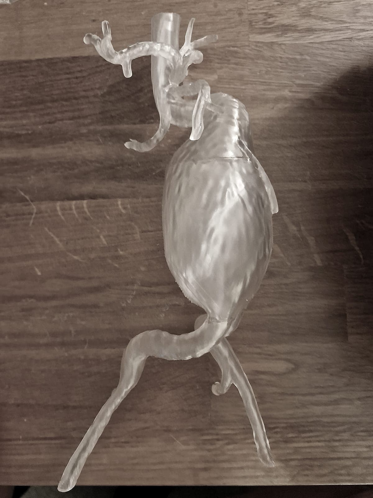
Clinician-led
Medical imaging and segmentation overseen by a clinical radiologist.
Patient-specific
Anatomical models derived directly from medical imaging datasets.
High-resolution
Detailed resin models produced for education and research.
Process
From scan to physical model
- 01
Define the project
We discuss the anatomy, structures and educational or research question.
- 02
Segment the imaging
The relevant anatomy is segmented from appropriately de-identified medical imaging.
- 03
Create the model
The segmentation is converted into a printable three-dimensional model and prepared for high-resolution resin printing.
- 04
Print & deliver
Models are printed, post-processed and delivered ready for their intended educational or research use.
Medical education research
Vascular anatomy education
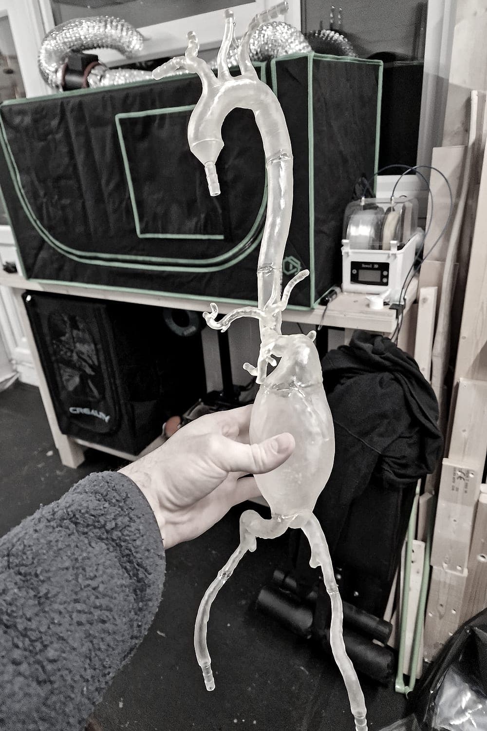
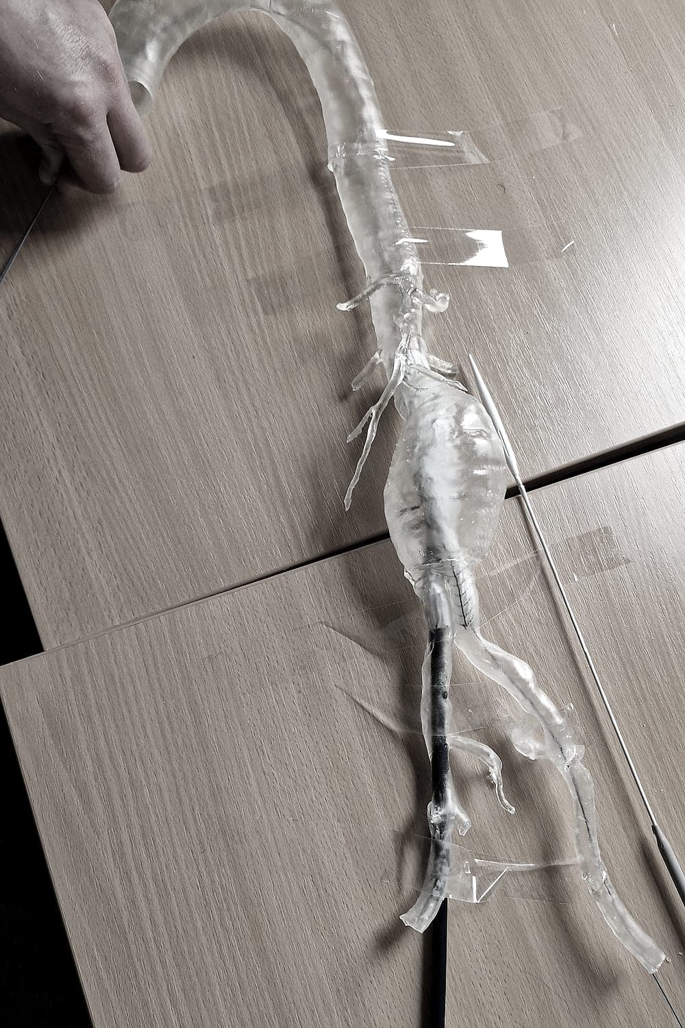
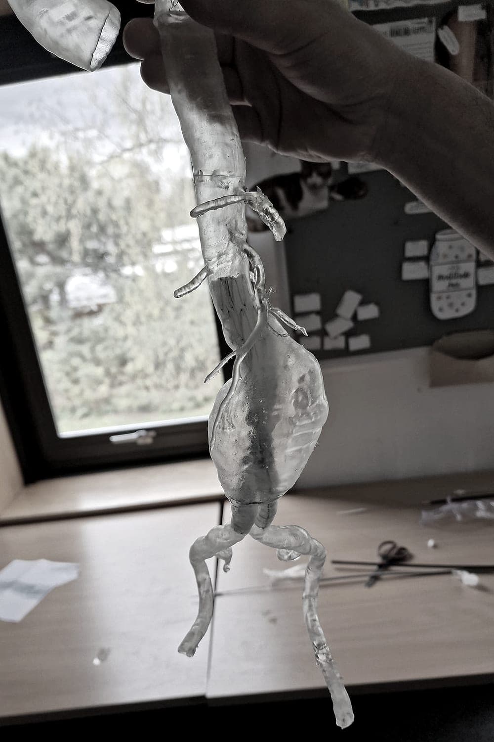
Eight patient-specific vascular anatomical models were produced for a postgraduate research project at a UK teaching hospital. The study is investigating whether physical 3D models can help medical students understand vascular anatomical variation.
The study is ongoing and findings are not yet available.
Applications
Built for medical education and research
Anatomy teaching
Patient-specific examples of anatomy and anatomical variation.
Medical education research
Physical anatomical models for studies evaluating novel approaches to teaching and learning.
Postgraduate training
Models for specialist teaching, demonstrations and workshops.
Radiology education
Transform complex cross-sectional imaging into tangible anatomy.
Simulation & skills training
Physical anatomical models for educational exercises and simulation environments.
Research projects
Bespoke model creation for academic and clinical research teams.
Why clinician-led matters
More than a 3D printing service
Producing a useful anatomical model starts long before the printer. Understanding the imaging, identifying the relevant anatomy and deciding what should, and should not, be included in the segmentation are fundamental to the final model.
- Clinical understanding of cross-sectional imaging
- Direct discussion with clinicians, researchers and educators
- Understanding of anatomical structures and variants
- Medical image segmentation expertise
- End-to-end ownership from imaging to physical model
Exploring a project involving 3D anatomical models?
Discuss a project
Founder, Health3D — clinical radiologist and machine-learning engineer.
About
Clinician-led medical 3D printing
Health3D was founded by a UK clinical radiologist and machine-learning engineer with experience across medical imaging, image segmentation and applied healthcare technology.
It began after being commissioned to produce a series of patient-specific vascular anatomical models for a medical education research project. That project highlighted a broader problem: healthcare researchers and educators may have access to medical imaging, but often not to the complete workflow required to:
- understand the imaging
- segment the anatomy
- prepare the model
- print it
- post-process it
- deliver a finished physical anatomical model
Health3D provides that end-to-end service.
FAQ
Common questions
What imaging can Health3D work from?
Primarily CT and other volumetric medical imaging suitable for three-dimensional segmentation. Projects can be discussed individually.
What information do you need?
The anatomy required, intended educational or research use, number of models and approximate timeline.
Can you produce multiple copies?
Yes. Once a digital model has been prepared, multiple physical copies can be produced.
How long does a project take?
Turnaround depends on segmentation complexity, model size and quantity. Contact Health3D to discuss individual project requirements.
How much does it cost?
Projects are currently quoted individually based on segmentation complexity, model size, printing requirements and quantity.
Can I upload medical imaging through this website?
No. Medical imaging and patient-identifiable data should not be submitted through the public website. Appropriate secure transfer arrangements can be discussed separately if a project proceeds.
Are Health3D models intended for clinical decision-making?
The current service is focused on models produced for education and research. Models are not currently supplied for diagnostic or clinical decision-making use.
Contact
Have a project in mind?
If you’re exploring the use of patient-specific anatomical models for teaching or research, I’d be happy to discuss what you’re trying to achieve and whether 3D printing is suitable.
Prefer email? enquiries@health3d.co.uk